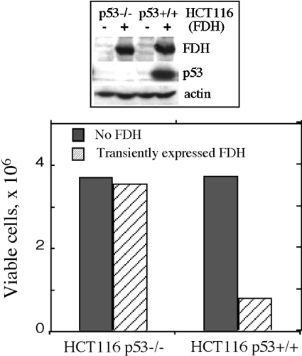
Figure 5. Effect of transient FDH expression on proliferation of p53 functional (HCT116 p53+/+) and p53-null (HCT116 p53−/−) cells.
Viable cells were assessed by Trypan Blue exclusion 48 h post-transfection. Cells were transfected with pcDNA 3.1/FDH vector using Lipofectamine 2000 (hatched bars). Control cells (solid bars, no FDH) were mock-transfected with ‘empty’ vector (no FDH cDNA insert). Efficiency of transfection evaluated by co-transfection with a vector expressing GFP was approx. 35%. Addition of geneticin allowed elimination of non-transfected cells, increasing the accuracy of this approach [29]. Upper panel: FDH levels in control (−) and FDH-transfected (+) cells detected by immunoblot assays.

