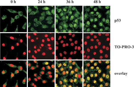
Figure 6. Accumulation of p53 in nuclei after induction of FDH expression in A549 cells.
Top row, p53 (green) was detected by immunostaining with monoclonal anti-p53 antibodies (clone DO-1, Calbiochem) and polyclonal anti-mouse IgG2a antibodies conjugated with Alexa Fluor 488 dye (Molecular Probes). Middle row, the nuclei (red) were imaged by chromosomal DNA staining with TO-PRO-3 iodide dye (Molecular Probes). Bottom row, overlay with yellow indicating co-localization. Images were obtained by confocal laser microscopy. Time (h) after induction of FDH expression is shown.

