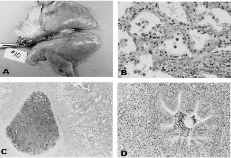
FIG. 7.
Pathological examination of the lungs of pigs 28 days after infection with KM22. (A) Gross photograph showing an area of tan, fibrous consolidation of the right cranial, middle, and part of the caudal lung lobes. (B) Photomicrograph showing moderate to marked thickening of the alveolar septa with fibrillar material, fibroblasts, and macrophages; marked type II pneumocyte hyperplasia and pneumocytes which contain abundant vacuolated cytoplasm; and alveolar lumina which contain variable quantities of fibrin, sloughed epithelium, and macrophages. Hematoxylin and eosin stain; 1 cm = 55 μm. (C) Photomicrograph showing abscessed lung lobule. Note the core of necrosis surrounded by a thick dense band of collagenous connective tissue. Hematoxylin and eosin stain. 1 cm = 350 μm. (D) Photomicrograph showing extensively focal obliteration of the alveolar lumina with infiltrates of large numbers of neutrophils and neutrophilic bronchiolitis. Note the infolding of the bronchiole mucosa and epithelial hyperplasia. Hematoxylin and eosin stain. 1 cm = 110 μm.

