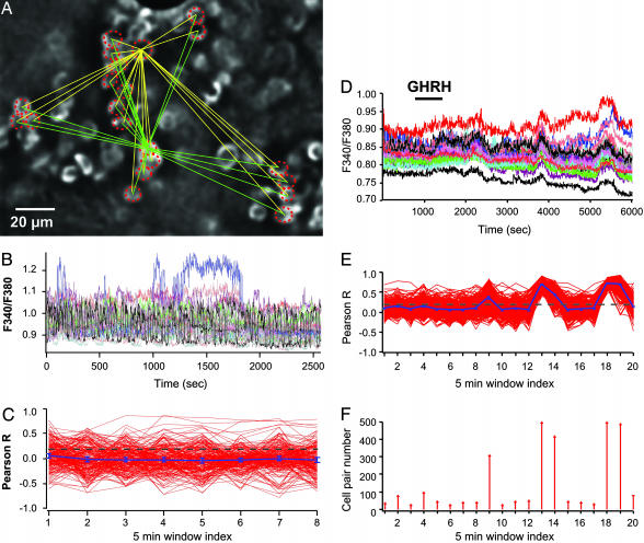
Fig. 4.
GHRH triggers recurrent motifs of GH cell connectivity in the lateral zones. (A) Field of GH cells labeled with EGFP in the lateral zone. Only cells circled with a dashed line were also loaded with the fluorescent calcium dye fura-2. (B) Representative traces of spontaneous calcium spikes due to electrical activity. (C) Linear correlation (Pearson R) between calcium recordings among all cell pairs (C1-C2, C1-C3,... CN-1-CN) taken every 5 min. The yellow and green lines (A) illustrate the potent cell pairs for two recorded GH cells. Although some cell pairs displayed high R values, no large-scale cell connectivity (P < 0.001) was observed during spontaneous calcium spiking. (D-F) GHRH (10 nM) was applied during a 15-min time period (horizontal bar). (D) GHRH triggered a complex, oscillating calcium response in GH-EGFP cells recorded in the lateral zone. (E) Recurrent motifs of connectivity among large cell ensembles were observed in lateral regions (P < 0.001). (F) Distribution of numbers of connected cell pairs vs. time of calcium recording. GHRH triggered a delayed, cycling increase in connected cell pairs.

