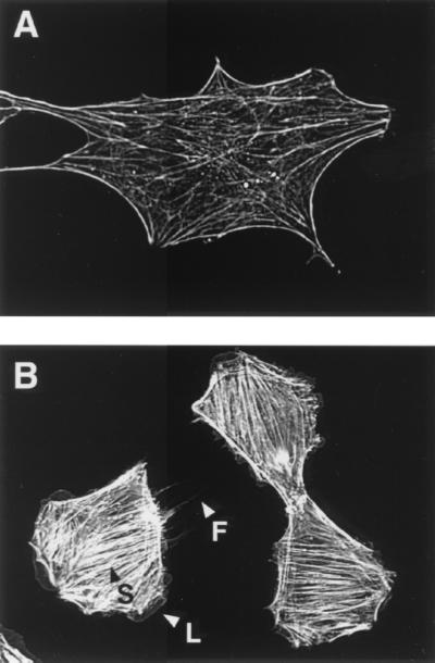
FIG. 4.
Formation of actin cytoskeletons in MC3T3-E1 cells treated with DNT. MC3T3-E1 cells in a subconfluent state were incubated in the presence (B) or absence (A) of DNT at 5 μg/ml for 24 h, and the actin fibers with rhodamine-phalloidin were observed under a fluorescence microscope. Arrowheads: S, stress fibers; L, lamellipodia; F, filopodia.

