Abstract
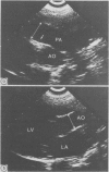
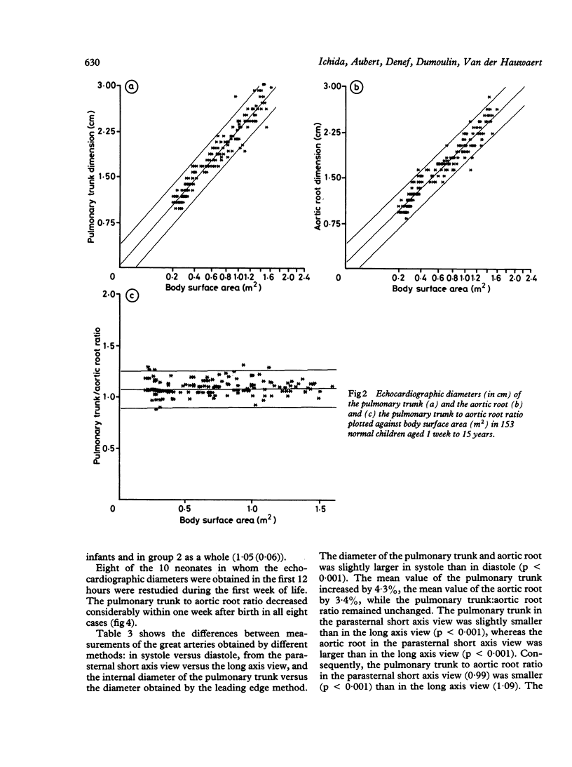
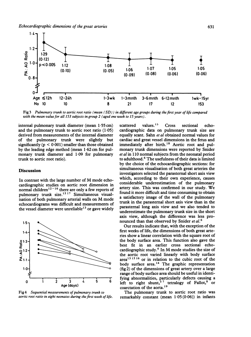
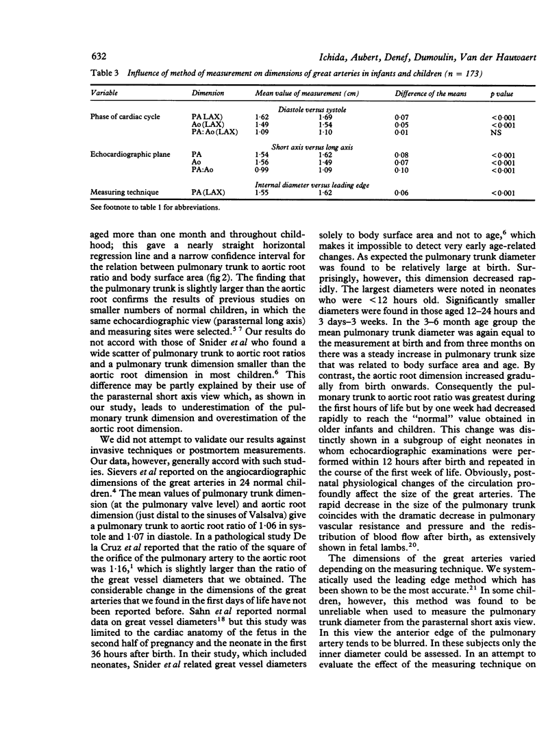
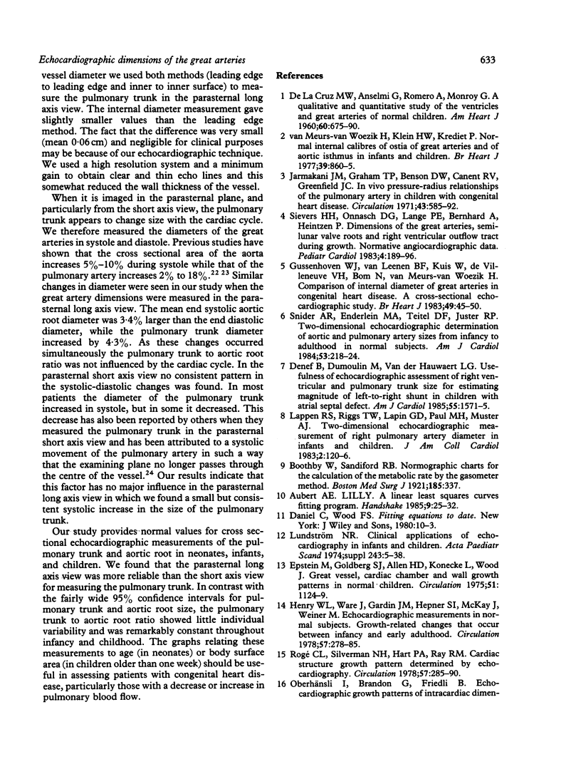
The pulmonary trunk and aortic root were measured on cross sectional echocardiograms in 173 normal subjects aged from one day to 15 years. Fifteen neonates were reexamined 3-6 days later. The great vessels were visualised in the parasternal long axis and short axis views. All measurements were made in end diastole and end systole by the leading edge method. The internal diameter (inner surface to inner surface) of the pulmonary trunk was also measured. The diameters of the great vessels correlated best with the square root of body surface area. Individual variability in cardiac growth gave a wide scatter of normal values. This was controlled for by calculating the ratio of the pulmonary trunk to aortic root for each subject. This ratio showed little individual variability and, except for the neonatal period, was remarkably constant throughout infancy and childhood (1.06 (0.06)). In the first 24 hours of life the ratio of the pulmonary trunk to the aortic root was significantly larger (1.29 (0.12)) but within one week it decreased to the "normal" ratio found in the older age groups. These normal data should be useful in assessing patients with congenital heart disease, particularly those in whom pulmonary blood flow is abnormal.
Full text
PDF

Images in this article
Selected References
These references are in PubMed. This may not be the complete list of references from this article.
- DE LA CRUZ M. V., ANSELMI G., ROMERO A., MONROY G. A qualitative and quantitative study of the ventricles and great vessels of normal children. Am Heart J. 1960 Nov;60:675–690. doi: 10.1016/0002-8703(60)90351-3. [DOI] [PubMed] [Google Scholar]
- Denef B., Dumoulin M., Van der Hauwaert L. G. Usefulness of echocardiographic assessment of right ventricular and pulmonary trunk size for estimating magnitude of left-to-right shunt in children with atrial septal defect. Am J Cardiol. 1985 Jun 1;55(13 Pt 1):1571–1575. doi: 10.1016/0002-9149(85)90975-0. [DOI] [PubMed] [Google Scholar]
- Epstein M. L., Goldberg S. J., Allen H. D., Konecke L., Wood J. Great vessel, cardiac chamber, and wall growth patterns in normal children. Circulation. 1975 Jun;51(6):1124–1129. doi: 10.1161/01.cir.51.6.1124. [DOI] [PubMed] [Google Scholar]
- GREENFIELD J. C., Jr, PATEL D. J. Relation between pressure and diameter in the ascending aorta of man. Circ Res. 1962 May;10:778–781. doi: 10.1161/01.res.10.5.778. [DOI] [PubMed] [Google Scholar]
- Gussenhoven W. J., van Leenen B. F., Kuis W., de Villeneuve V. H., Bom N., van Meurs-van Woezik H. Comparison of internal diameter of great arteries in congenital heart disease. A cross-sectional echocardiographic study. Br Heart J. 1983 Jan;49(1):45–50. doi: 10.1136/hrt.49.1.45. [DOI] [PMC free article] [PubMed] [Google Scholar]
- Hagan A. D., Deely W. J., Sahn D., Friedman W. F. Echocardiographic criteria for normal newborn infants. Circulation. 1973 Dec;48(6):1221–1226. doi: 10.1161/01.cir.48.6.1221. [DOI] [PubMed] [Google Scholar]
- Henry W. L., Ware J., Gardin J. M., Hepner S. I., McKay J., Weiner M. Echocardiographic measurements in normal subjects. Growth-related changes that occur between infancy and early adulthood. Circulation. 1978 Feb;57(2):278–285. doi: 10.1161/01.cir.57.2.278. [DOI] [PubMed] [Google Scholar]
- Jarmakani J. M., Graham T. P., Jr, Benson D. W., Jr, Canent R. V., Jr, Greenfield J. C., Jr In vivo pressure-radius relationships of the pulmonary artery in children with congenital heart disease. Circulation. 1971 Apr;43(4):585–592. doi: 10.1161/01.cir.43.4.585. [DOI] [PubMed] [Google Scholar]
- Morrow W. R., Huhta J. C., Murphy D. J., Jr, McNamara D. G. Quantitative morphology of the aortic arch in neonatal coarctation. J Am Coll Cardiol. 1986 Sep;8(3):616–620. doi: 10.1016/s0735-1097(86)80191-7. [DOI] [PubMed] [Google Scholar]
- Oberhänsli I., Brandon G., Friedli B. Echocardiographic growth patterns of intracardiac dimensions and determination of function indices during the first year of life. Helv Paediatr Acta. 1981 Sep;36(4):325–340. [PubMed] [Google Scholar]
- Rogé C. L., Silverman N. H., Hart P. A., Ray R. M. Cardiac structure growth pattern determined by echocardiography. Circulation. 1978 Feb;57(2):285–290. doi: 10.1161/01.cir.57.2.285. [DOI] [PubMed] [Google Scholar]
- Sahn D. J., Lange L. W., Allen H. D., Goldberg S. J., Anderson C., Giles H., Haber K. Quantitative real-time cross-sectional echocardiography in the developing normal humam fetus and newborn. Circulation. 1980 Sep;62(3):588–597. doi: 10.1161/01.cir.62.3.588. [DOI] [PubMed] [Google Scholar]
- Sievers H. H., Onnasch D. G., Lange P. E., Bernhard A., Heintzen P. H. Dimensions of the great arteries, semilunar valve roots, and right ventricular outflow tract during growth: normative angiocardiographic data. Pediatr Cardiol. 1983 Jul-Sep;4(3):189–196. doi: 10.1007/BF02242254. [DOI] [PubMed] [Google Scholar]
- Snider A. R., Enderlein M. A., Teitel D. F., Juster R. P. Two-dimensional echocardiographic determination of aortic and pulmonary artery sizes from infancy to adulthood in normal subjects. Am J Cardiol. 1984 Jan 1;53(1):218–224. doi: 10.1016/0002-9149(84)90715-x. [DOI] [PubMed] [Google Scholar]
- van Meurs-Van Woezik H., Klein H. W., Krediet P. Normal internal calibres of ostia of great arteries and of aortic isthmus in infants and children. Br Heart J. 1977 Aug;39(8):860–865. doi: 10.1136/hrt.39.8.860. [DOI] [PMC free article] [PubMed] [Google Scholar]


