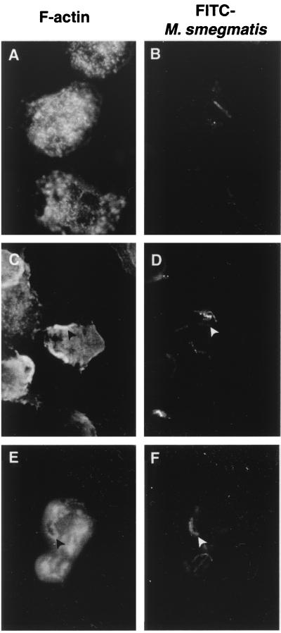
FIG. 5.
Phagocytosis of mycobacterial aggregates induces actin polymerization. Adherent neutrophils were incubated in the presence of unicellular FITC-stained mycobacteria (50:1) for 30 min (A and B) or aggregated FITC-stained mycobacteria (C to F) for 5 min (C and D) or 30 min (E and F). Cells were fixed and permeabilized in acetone at −20°C, and F-actin was stained with rhodamine-conjugated phalloidin (A, C, and E) visualized by fluorescence microscopy. At 5 min, phagocytic cups of F-actin (C) (black arrowhead) are seen in front of a mycobacterial clump (D) (white arrowhead). At 30 min p.i., phagosomes containing mycobacterial aggregates are surrounded by F-actin (E and F), whereas this is not seen around phagosomes containing unicellular mycobacteria (A and B). One representative experiment out of three is shown.

