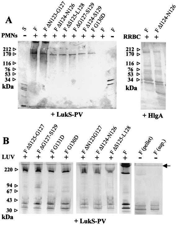
FIG. 4.
Characterization of leucotoxin oligomers on different membrane systems. (A) Human PMNs or RRBC (30 × 106 cells/ml) were treated with 60 nM S protein and 600 nM F protein before being submitted to SDS-PAGE, and oligomers were revealed by immunoblotting with both anti-LukS-PV and anti-LukF-PV rabbit polyclonal antibodies. Combinations of LukF-PV or mutants were tested with either LukS-PV or HlgA. Controls of cells with components alone or of components with (+) and without (−) cells are indicated. (B) Assembly of leucotoxin oligomers by native and mutated LukF-PV on model membranes. Leucotoxin oligomers formed by the indicated form of LukF-PV in association with either LukS-PV were detected by SDS-PAGE of the vesicles pelleted after ultracentrifugation. Controls of native leucotoxin without lipids, separated in pellet and supernatant, are included in the left panel. Some streaking appears in the lanes with lipids. The end of the stacking gel is indicated by an arrow. Left lane, molecular standards.

