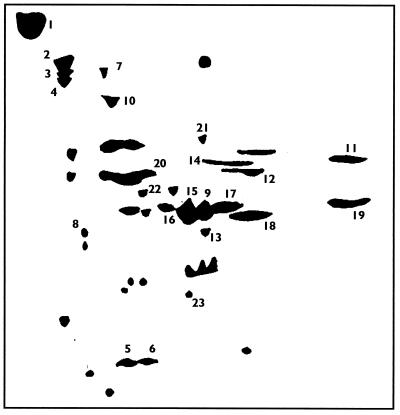
FIG. 1.
Representation of the outer surface proteins of S. agalactiae M732 separated by 2-D gel electrophoresis. Proteins were isolated from the outer surface of S. agalactiae by enzymatic digestion. Following concentration and dialysis, samples of protein (300 μg to 1 g) were analyzed through the first dimension over a pH range of 3 to 10 prior to application on a second-dimension SDS-10% polyacrylamide gel. The gel was stained using GelCode blue (Pierce). The representation is a compilation of gels which were loaded with differing amounts of protein.

