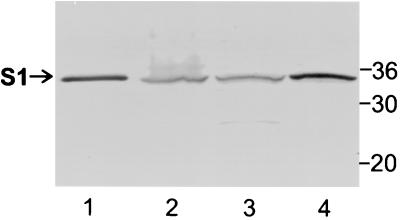
FIG. 2.
Immunoblot analysis of the S1 subunit in cell fractions of wild-type strain B. pertussis BP536. The cell fractionation procedures used and the methods used to prepare samples are described in Materials and Methods. Samples were subjected to SDS-PAGE and immunoblot analysis using monoclonal antibody 3CX4 to visualize the S1 subunit of pertussis toxin. Equivalent amounts of the cytoplasmic-periplasmic fraction and the total membrane fraction (inner and outer membranes) were loaded. Six times more of each of these fractions than whole-cell extract was loaded. The positions of molecular mass markers (in kilodaltons) are indicated on the right. The arrow indicates the protein band corresponding to full-length S1. Lane 1, purified pertussis toxin (0.1 μg); lane 2, whole-cell extract; lane 3, cytoplasmic-periplasmic fraction; lane 4, total membrane fraction.

