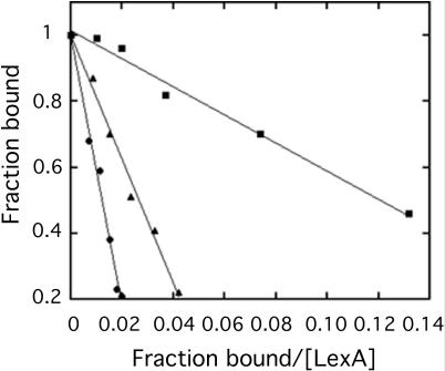
Figure 3.
Representative Scatchard plots for quantifying inhibition of LexA binding to the recA operator by operator mutants. Mobility shift assays were conducted as described in Materials and Methods with purified LexA, radiolabeled recA promoter DNA (5.0 nM), and a 100-fold molar excess of wild-type recA operator (black circles), recA operator with A substituted for C at the first position (black triangles) and no competitor (black squares). LexA concentrations used were for unbound LexA.

