Figure 4.
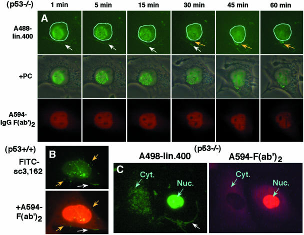
Movement to the cytoplasm of DNA injected into the nucleus. (A) Alexa 488-labeled lin. 400 DNA was mixed with Alexa 594-labeled IgG F(ab′)2 protein, and co-injected into the nucleus of MEF p53−/− cells. Within 1 min, the injected DNA appeared at the periphery of the cytoplasm (white arrows). Furthermore, a portion of the aggregates that were formed within the nucleus moved to the cytoplasm: this was obvious at 30, 45 and 60 min (yellow arrows). In the fluorescence images, the nucleus rim was outlined with cyan lines. (B) FITC-labeled sc3,162 DNA was injected into the nucleus of MEF p53+/+ cells and the cells were fixed 30 min later. A portion of the DNA was detected at either the cytoplasmic rim (white arrow) or as an aggregate in the cytoplasm (yellow arrow). (C) The cells were injected as described in (A). If the DNA was injected into the nucleus (Nuc.), a portion of it was detected at the cytoplasmic rim at 3 min after the injection (white arrow), whereas direct injection into the cytoplasm (Cyt.) did not lead to this distribution.

