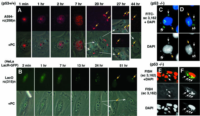
Figure 6.
Stabilization of intranuclear-injected DNA in the nucleus and its localization in the cytoplasm after mitosis. Alexa 594-labeled rc(258)n (A) or LacO rc(315)n DNA (B) was injected into the nucleus of MEF p53−/−(A) or HeLa LacR-GFP (B) cells. The injected DNA aggregated in the nucleus (A and B) and the number of aggregates decreased as time progressed (B). After mitosis, the aggregated DNA appeared in the cytoplasm near the nucleus [(A and B); yellow arrows). Such cytoplasmic aggregates were also detected where FITC-labeled sc 3,162 DNA was injected into the nucleus of MEF p53−/− cells and fixed with paraformaldehyde at 24 h (C, D). Furthermore, unlabeled sc 3,162 (pUC119) DNA was injected into the nucleus of MEF p53−/− cells and fixed with paraformaldehyde at 24 h (E and F). The injected DNA was detected by FISH using a probe prepared from pUC119 DNA. The cellular DNA was counter-stained with DAPI (C and D) or propidium iodide (E and F). In this case as well, the DNA was detected as aggregates in the cytoplasm. In (C–F), higher concentrations of DNA [200 ng/µl in (C and D), and 500 ng/µl in (E and F)] were injected compared with the normal conditions employed in this study. In these cases, the cytoplasmic aggregate (white arrows) looked just like micronucleus that was spontaneously formed in these cells (yellow arrows).

