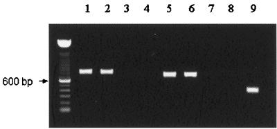
FIG. 1.
RT-PCR analysis of total RNA extracted from B. bovis-infected erythrocytes (lanes 1 and 5), B. bovis-infected larvae (lanes 2 and 6), B. bovis-infected larvae without RT treatment (lanes 3 and 7), and uninfected larvae (lanes 4, 8, and 9). Amplification used primers specific for rap-1 (lanes 1 to 4, 800 bp), msa-1 (lanes 5 to 8, 711 bp), and Bm86 (lane 9, 400 bp). Amplicons were detected by agarose gel electrophoresis and ethidium bromide staining. Molecular size markers are shown on the left.

