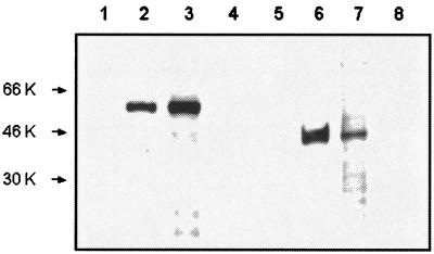
FIG. 2.
Immunoblot analysis of total protein extracted from normal erythrocytes (lanes 1 and 5), B. bovis-infected erythrocytes (lanes 2 and 6), B. bovis-infected B. microplus larvae (lanes 3 and 7), and uninfected B. microplus larvae (lanes 4 and 8). Protein extracts were electrophoresed on sodium dodecyl sulfate-containing polyacrylamide gels and transferred to nitrocellulose membranes. The membranes were incubated with MAb BABB75A4 against RAP-1 (lanes 1 to 4) or MAb 23/10.36.18 against MSA-1 (lanes 5 to 8). Molecular size markers are on the left.

