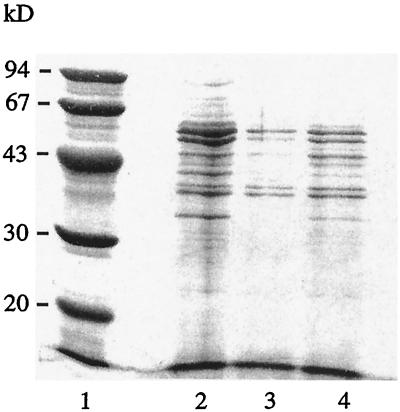
FIG. 1.
Electrophoretic analysis on SDS-12% PAGE with Coomassie brilliant blue staining. Shown are molecular mass markers (lane 1), Salmonella antigens from pooled cultures before conjugation to the microparticles (lane 2), Salmonella antigens from the supernatant remaining after the conjugation (lane 3), and Salmonella antigens from a single culture (lane 4).

