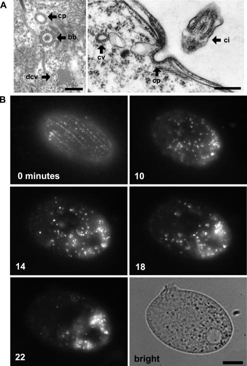
Figure 2. Visualizing Sites of Endocytosis.
(A) In Tetrahymena, coated pits (cp) are found near the base of cilia, as shown in tangential (left) and cross (right) sections. bb, ciliary basal body; ci, cilia; cv, coated vesicle; mt, mitochondrion; dcv, dense core vesicle. Bars = 200 nm.
(B) Time course of FM1–43 dye uptake. A cell shown immediately after treatment with 5 μM FM1–43 (0 min) shows rows of fluorescent puncta at the cell surface. Time-lapse images (10, 14, 18, and 22 min) of a single cell following 5-min exposure to FM1–43. At the later time points (18 and 22 min), the dye accumulates in what appear as vesicles clustered toward the cell posterior. The brightfield image shows the cell at the end of the time course. Bar = 10 μm.

