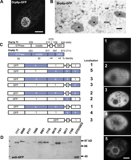
Figure 10. Analysis of Drp1p Targeting by Chimera Analysis.
(A) Compiled stack of confocal sections through fixed cell expressing Drp6p-GFP (driven by the MTT1 promoter) shows localization at the nuclear envelope. Bar = 10 μm.
(B) Immuno-gold visualization of Drp6p-GFP shows localization in clusters on the cytoplasmic face of the nuclear envelope (ne). Regions of heterochromatin within the macronucleus (mac) are indicated (*). Bar = 200 nm.
(C) Domain comparison of Drp1p and Drp6p and diagrams of Drp-GFP chimeras (expressed under the MTT1 promoter) indicating localization. Numbered images at right show localization patterns observed in cells expressing chimeric proteins. Bar = 10 μm. nd, not detected.
(D) Western blot of total cell lysates from Drp-GFP cells confirms stability of full-length chimeras.

