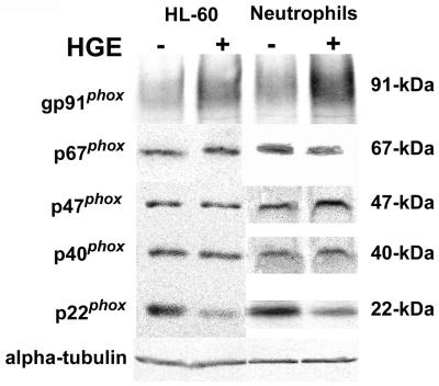
FIG. 1.
Analysis of cellular levels of NADPH oxidase components following exposure to A. phagocytophila. HL-60 culture cells or human neutrophils were infected with A. phagocytophila (HGE) for 7 days or 30 min, respectively. Cell lysates (25 μg) of infected and uninfected cells were subjected to SDS-PAGE and Western immunoblotting and probed with antibodies against gp91phox (rabbit), p67phox, p47phox, p40phox, p22phox, and α-tubulin. Results shown are representative of three independent experiments.

