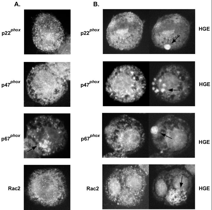
FIG. 3.
Double immunofluorescence labeling of the NADPH oxidase enzyme components p22phox, p47phox, p67phox, and Rac2, which appear as white dots in uninfected (A) or A. phagocytophila-infected (B) HL-60 cells. Note reduced amounts of p22phox in infected cells compared to those in uninfected cells. An arrow in the panel showing p67phox labeling in uninfected HL-60 cells (A) indicates an area of intense fluorescent labeling near the Golgi apparatus. Arrows in the infected HL-60 cells (B) indicate the locations of A. phagocytophila (HGE) morulae. Magnification, ×750. Results are representative of three independent labeling experiments.

