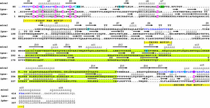
Fig. 2.
Alignment of the sequences of MICALfd with structural homologs. Single-letter amino acid codes are used. Three sequences are included. (i) MICAL: residues 85-442 of the MICAL-1 of mouse. Residues 1-484 of this protein correspond to MICALfd, but residues 1-84 of MICALfd form a domain that is not present in the other enzymes. (ii) Human monoamine oxidase B (Protein Data Bank entry 1GOS). Only residues 4-95 and 198-456 are shown. (iii) pHBH from Pseudomonas fluorescens residues 1-340 (Protein Data Bank entry 1PBE). The positions of the elements of secondary structure of MICAL, pHBH, and 1GOS are shown at the top of each sequence. The sequences corresponding to the conserved motifs are shown with blue letters. Letters with green background correspond to regions with large insertions and deletions. The two alanine residues with the green background correspond to the mutations introduced to improve the crystals. Residues identical in all three sequences have magenta backgrounds. Residues that contact the FAD cofactor are shown with cyan background. An insertion of 100 residues between α7 and β4, present only in amine oxidase, has been omitted.

