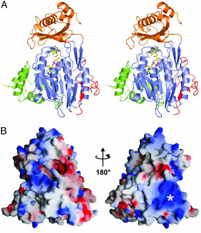
Fig. 1.
Crystal structure of mMICAL489. (A) Stereoview of mMICAL489 with four-helix bundle domain (1-85, green), FAD-binding domain (86-234 and 367-444, slate), MO domain (235-366, orange) and linker region (445-489, red) shown. The FAD molecule is depicted as sticks. (B) Solvent-accessible surface of mMICAL489 colored by electrostatic potential contoured at ±15 kT using grasp (36) (red, acidic; blue, basic). Left-hand view is shown, as in A. The asterisk marks a patch of basic potential.

