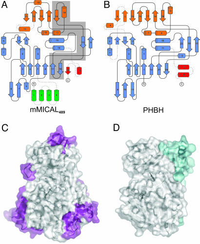
Fig. 2.
Structural comparison of mMICAL489 and PHBH. (A) Topology of mMICAL489 (β-strands, arrows; α-helices, cylinders). Domains are colored as in Fig. 1 A. Dotted lines denote unique structural elements. The gray-shaded area is deleted in the human splice isoform MICAL-1B (6); this deletion appears to be incompatible with formation of a stable molecule. (B) Equivalent diagram for PHBH. (C) Solvent-accessible surface of mMICAL489 with parts unique to mMICAL489 (compared with PHBH) highlighted in violet (orientation is as in Fig. 1 A). (D) Solvent-accessible surface of PHBH (oriented to superpose on mMICAL489) with parts unique to PHBH (compared with mMICAL489) highlighted in cyan.

