FIG. 1.
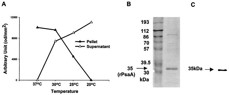
Expression of fusion protein (rPsaA-intein-CBD) (A), identification of the purified rPsaA protein by SDS-PAGE (B), and Western blot analysis (C). (A) Fusion proteins were expressed in E. coli XL1-Blue at 37, 30, 25, or 20°C. Insoluble and soluble proteins were recovered in pellets (▴) and supernatants (▵), respectively. The density of the fusion protein band was determined with TINA software and plotted against temperature. od, optical density units. (B and C) After purification, the rPsaA solution was separated on 10% polyacrylamide gels (B) and probed with sera from rabbits immunized with S. pneumoniae (C). The numbers in panel B indicate molecular masses of marker proteins. An arrow indicates the purified rPsaA protein (35 kDa).

