FIG. 5.
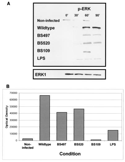
Time course of ERK phosphorylation by S. flexneri. T84 cells were exposed to wild-type S. flexneri or the LPS mutant strains at an MOI of 400 for various times prior to preparation of cellular lysates. (A) Western blot performed with specific antibodies for dually phosphorylated ERK1/2. Incubation with HBSS+ buffer served as the uninfected control. Equal loading of protein was confirmed by probing the immunoblots for ERK1. (B) Optical density tracing of phosphorylated ERK following a 90-min infection of T84 cells by S. flexneri. The data shown represent one of at least three experiments performed with similar results.

