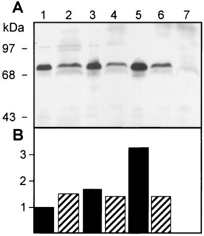
FIG. 1.
(A) Western blot analysis of dnaK expression in total cell lysates of wild-type B. suis (lanes 1, 3, and 5) and the dnaKmut strain (lanes 2, 4, and 6) with monoclonal anti-DnaK antibody. Growth temperatures were 30°C (lanes 1, 2, and 7), 37°C (lanes 3 and 4), and 46°C (lanes 5 and 6). The dnaK null mutant was used as a control (lane 7). (B) Quantification of DnaK in panel A, using SigmaGel software, relative to the amount of DnaK present in the wild-type lysate at 30°C (lane 1).

