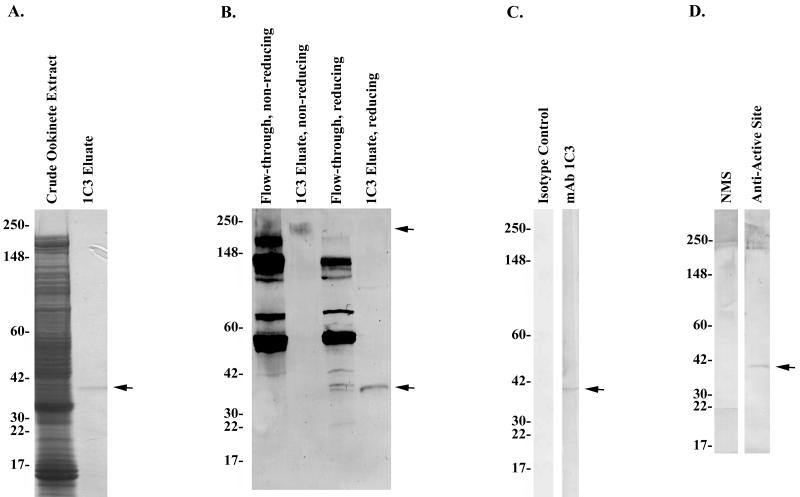
FIG. 6.
Analysis of MAb 1C3 affinity column-isolated P. gallinaceum antigen. (A) Coomassie blue-stained SDS-4 to 20% PAGE gel of P. gallinaceum ookinete extract or supernatant column starting material (lane 1) and 1C3 affinity-isolated ∼35-kDa antigen (lane 2). The arrow indicates the MAb 1C3-isolated protein. (B) MAb 1C3 Western immunoblot of flowthrough fractions or MAb 1C3 affinity-isolated proteins from 1C3 affinity chromatography. Western blots were performed after the proteins were separated under either nonreducing or reducing SDS-PAGE conditions as noted above the membrane. Arrows indicate the isolated proteins analyzed under either reducing or nonreducing SDS-PAGE conditions. (C) Western immunoblot recognition of isolated P. gallinaceum antigens by isotype control MAb (lane 1) and MAb 1C3 (lane 2). (D) Identification of 1C3 epitope-containing protein as cross-reactive with an antiserum raised against a peptide derived from the conserved Plasmodium chitinase active site (29). A total of 5 μg of P. gallinaceum ookinete extract and/or supernatants and 8 μg of 1C3 affinity-isolated ookinete antigens was loaded per lane, respectively. NMS, normal mouse serum.

