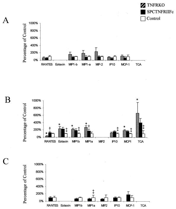
FIG. 5.
Altered chemokine expression in the lungs of SPCTNFRIIFc mice after aerosol infection with M. tuberculosis. Lung chemokine gene expression was determined by RNase protection assay of 20 μg of whole lung RNA from four animals per group (TNFRKO, SPCTNFRIIFc, and control mice) at 2 weeks (A), 3 weeks (B), and 4 weeks (C) after infection. Densitometry was performed on each specimen, and gene expression was normalized to internal standards (GADPH and L32) and then compared to the expression in control mice. Preliminary experiments comparing cytokine and chemokine expression levels from uninfected TNFRKO, SPCTNFRIIFc, and control mice demonstrated undetectable expression of all messages except for internal standards for RNA quantity. Chemokine values are expressed as percentages of expression in infected control mice, and error bars represent the standard deviations. Significant differences (P < 0.05) between TNFRKO and control mice are indicated by asterisks (∗), between TNFRKO and SPCTNFRIIFc mice by daggers (†), and between SPCTNFRIIFc and control mice by double daggers (‡).

