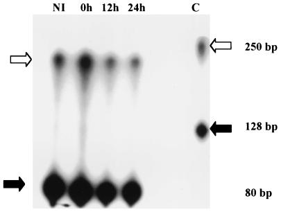
FIG. 3.
Results of RPA of bovine thymosin β-10. RNA from M. bovis-noninfected (NI) and -infected macrophages at 0, 12, and 24 h postinfection was hybridized to a thymosin β-10-radiolabeled probe, digested with RNase, and loaded into a 6% polyacrylamide gel. White arrows indicate the thymosin β-10-protected fragments on the right and the control probe (C) on the left, whereas the black arrows indicate the protected fragments of the ribosomal 18S internal control on the right and control probe on the left. Thymosin β-10 mRNA expression increased twofold after infection (0 h), while the level of expression in noninfected cells at 0 h was considered baseline. The fold increase was obtained as a ratio of the size and density of the protected fragment representing the differentially expressed gene to the size and density of the protected fragment of the internal control. Values are representative of two independent experiments.

