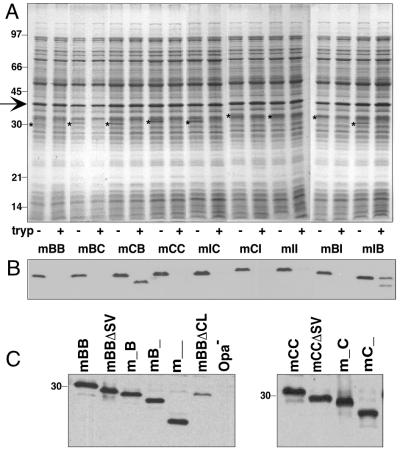
FIG. 3.
Expression of deletion and chimeric Opa proteins in gonococcal strain MS11. (A) Expression and surface exposure of chimeric Opa proteins determined by limited trypsinolysis. Whole bacteria were exposed to trypsin as described in Materials and Methods. Cell lysates were analyzed by SDS-PAGE and stained with Coomassie brilliant blue. Molecular mass standards are indicated on the left in kilodaltons. Arrow, porin protein; *, Opa proteins; − and +, absence or presence of trypsin (tryp), respectively. (B) Detection of Opa proteins by immunoblotting with anti-Opa antibody 4B12. Samples are identical to those shown in the gel in panel A. (C) Expression of deletion Opa variants. Whole-cell lysates were separated by SDS-PAGE, and Opa proteins were detected by immunoblotting with anti-Opa antibody 4B12. _, deletions of HV regions (i.e., m_B = mBB with its HV-1 loop deleted; mB_ = mBB with its HV-2 loop deleted).

