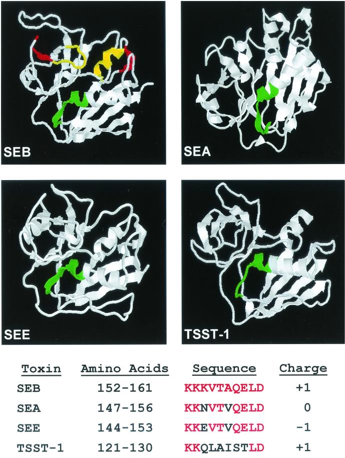
FIG. 4.
Molecular models of SEB, SEA, SEE, and TSST-1. The amino acids highlighted in green signify the conserved sequence (KKKVTAQELD in SEB). The yellow amino acids on SEB identify the areas implicated in MHC-II binding, and the red amino acids correspond to the binding site of the TCR. All structures were obtained from the Protein Data Bank of the Research Collaboratory for Structural Bioinformatics. Downloaded files were subsequently manipulated using the RasMol program. The coordinates are based upon original publications about SEB (25), SEA (29), SEE (34), and TSST-1 (26).

