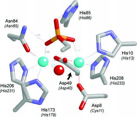
Figure 10.
Proposed similarity of the active sites of Mre11 and Dbr1. The Figure depicts the active site of the manganese- and 5′-dAMP-bound Pyrococcus furiosus phosphodiesterase Mre11 from the crystal structure (PDB 1II7). The amino acid side chains coordinating the binuclear metal cluster and the nucleotide 5′-phosphate are shown. The corresponding amino acids of yeast Dbr1 are indicated in parentheses. The manganese ions are colored cyan. Water is colored red. For simplicity, only the phosphate and the ribose C4 and C5 atoms of the nucleotide are shown.

