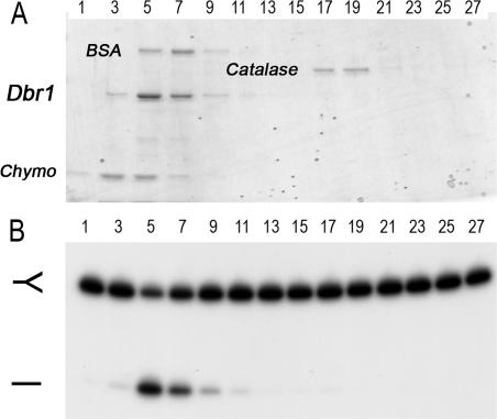
Figure 8.
Glycerol gradient sedimentation of Dbr1. Sedimentation was performed as described under Materials and Methods. (A) Aliquots (9 µl) of the odd-numbered gradient fractions were analyzed by SDS–PAGE. The Coomassie blue-stained gel is shown. The positions of the recombinant Dbr1 and the marker proteins chymotrypsinogen (Chymo), BSA and catalase are indicated. (B) Reaction mixtures (20 µl) containing 5 nM of 32P-labeled branched oligonucleotide substrate (depicted in Figure 6B) and 2 µl of a 1:100 dilution of the odd-numbered glycerol gradient fractions were incubated for 10 min at 22°C. The products were analyzed by denaturing PAGE and visualized by autoradiography.

