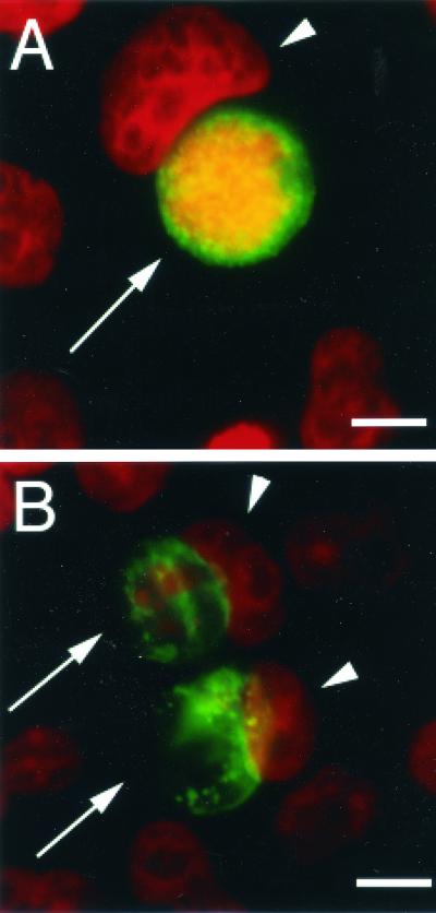
FIG. 2.
Appearance of Chlamydia inclusions in cells treated with IFN-γ. HeLa cells infected with C. muridarum and treated with IFN-γ at 0 (A) or 2 (B) ng/ml were fixed with paraformaldehyde and prepared for immunofluorescence as described in Materials and Methods, using fluorescein-conjugated anti-Chlamydia antibodies (green) and Hoechst for DNA labeling (red). Chlamydia inclusions were normal in the absence of IFN-γ but were aberrant when infected cells were treated with 2 ng of IFN-γ per ml. Arrows indicate Chlamydia inclusions, and arrowheads show host cell nuclei. Bar, 10 μm.

