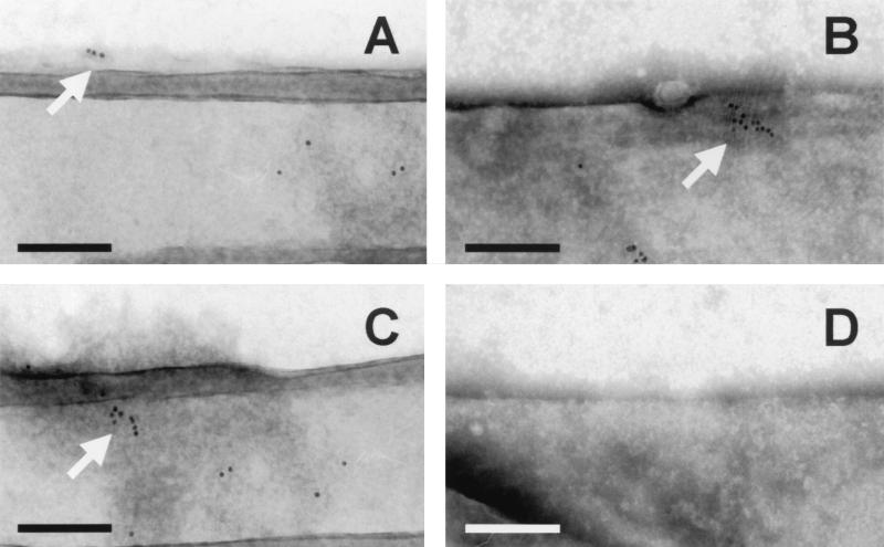
FIG. 5.
Immunoelectron microscopy of M. tuberculosis using MAb 24c5. M. tuberculosis Erdman grown in the absence of Tween 80 for 25 days was analyzed for binding of MAb 24c5 (A to C) by using goat anti-mouse IgG sera conjugated to 5-nm gold. Gold appears as black dots, as indicated by arrows (A to C). The images in panels A to C are from three different bacterial fields and two different M. tuberculosis samples. Irrelevant control antibody did not allow binding of gold-conjugated sera (D). Bars = 200 nm.

