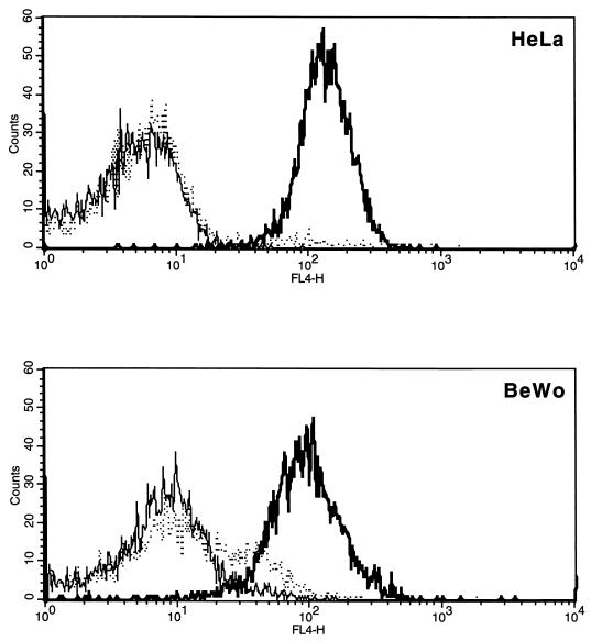
FIG. 3.
Flow cytometry analysis of IFN-γ receptor (CD119) expression on BeWo and HeLa cells. Cells were stained by a three-step procedure involving a primary mouse monoclonal antibody followed by an anti-mouse biotin conjugate and then streptavidin-allophycocyanin. Cells stained with a monoclonal antibody reacting with human CD119 are shown by the heavy solid line. The dotted line shows cells reacted with an isotype-matched control, and the light solid line shows cells stained with the second- and third-step reagents only.

