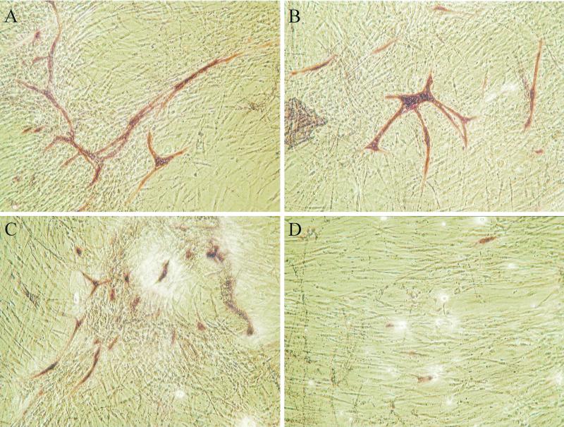
FIG. 4.
Impact of HdCDT on an in vitro model of angiogenesis. ECs and tubuli were visualized by staining for PECAM (CD31) (shown in red). In control (PBS-treated) wells long tubuli are formed (A). With 102 CPU (B) and 104 CPU (C) of HdCDT per ml the tubuli are shorter and less branched. Increasing the concentration further to 105 CPU HdCDT per ml (D) totally inhibits the formation of tubuli. The cell density of the fibroblast-like cells is decreased at high concentrations of HdCDT (D) compared to control (A).

