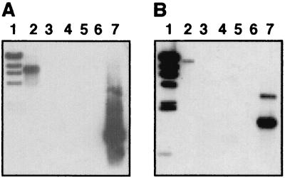
FIG. 2.
Southern blot analysis of H. influenzae biogroup aegyptius genomic and plasmid DNAs. Genomic DNAs from F3031 (lane 2) and F1947 (lane 3) and pF3031 (lane 4) were digested with HindIII. EcoRI-digested pMU34 and pMU33 were loaded into lanes A5 and B5, respectively. In both panels, lane 1 contained HindIII-digested λ DNA and lane 6 was empty. PCR amplicons of the pMU33 (lane A7) and pMU34 (lane B7) inserts were used as positive controls. DNA fragments were separated by agarose gel electrophoresis and blotted onto nitrocellulose. Blots were probed with 32P-labeled HindIII-digested λ DNA and 32P-labeled PCR amplicons of MU33 (A) and MU34 (B).

