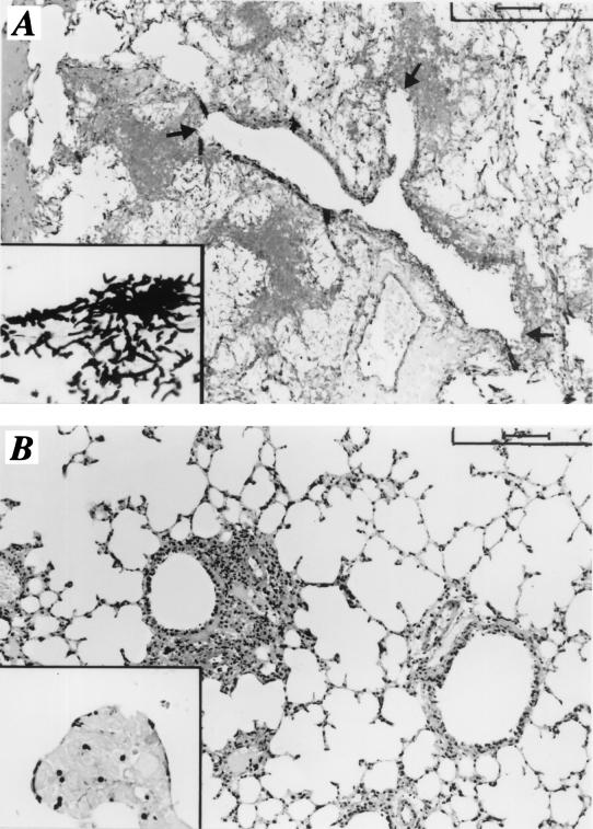
FIG. 6.
Histology of lungs of mice with IPA treated or not treated with the K10 MAb. Lethally irradiated C3H/HeJ mice received transplants of allogeneic T-cell-depleted BM cells from DBA/2 mice. Three days later, the mice were infected with 2 × 10⁷ A. fumigatus conidia, which were given intranasally for 3 consecutive days, and treated with a control MAb (A) or the K10 MAb (B). Periodic acid-Schiff (A and B)-stained or Gomori-Grocott (insets)-stained sections were prepared from the lungs of mice 1 day after the last intranasal infection. Note the presence of numerous hyphae and evident signs of bronchial wall destruction (arrows) in the lungs of control mice (A and inset), as opposed to the presence of a few swollen conidia in the lungs of mice treated with the K10 MAb (B and inset). Bars, 100 μm (A and B) and 25 μm (insets).

