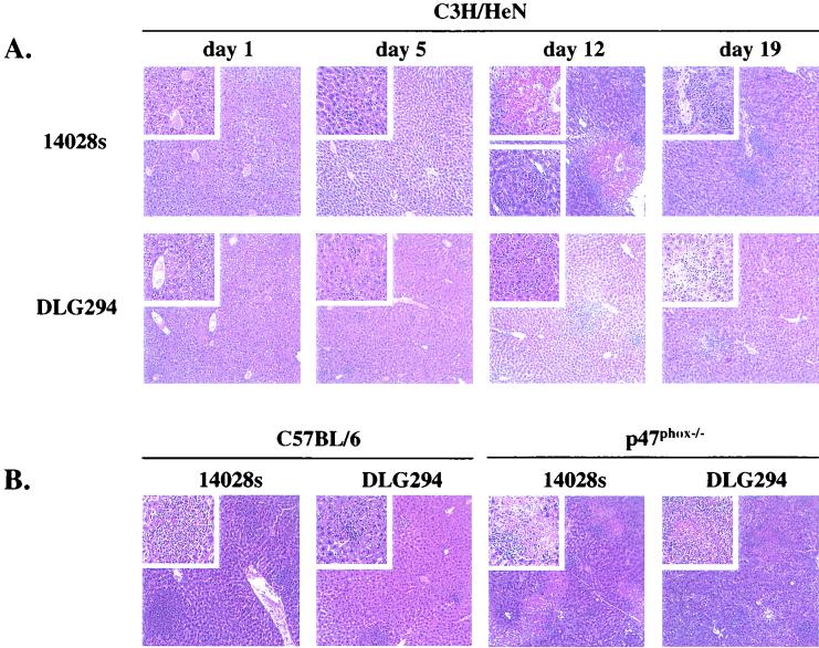
FIG. 2.
Microscopic appearance of livers from C3H/HeN mice infected with S. enterica serovar Typhimurium 14028s and DLG294 at several time points after infection (A) and C57BL/6 and p47phox−/− mice at 4 days after infection with S. enterica serovar Typhimurium 14028s and DLG294 (B). Mice were infected subcutaneously in the flanks as described in Materials and Methods. hematoxylin- and eosin-stained sections were prepared from livers that were isolated at different time points after infection.

