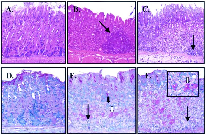
FIG. 2.
Histochemical analysis of tissue extracts from H. pylori- or A. lwoffii-inoculated mice. H&E staining of tissue extracts from control (A) and H. pylori (B)- and A. lwoffii (C)-inoculated mice shows significant inflammation as indicated by the arrows. PAS-alcian blue staining of tissue extracts from control (D) and H. pylori (E)- and A. lwoffii (F)-inoculated mice shows neutral (white arrow) and acid (solid arrow) mucins. Magnification, ×400 (all panels).

