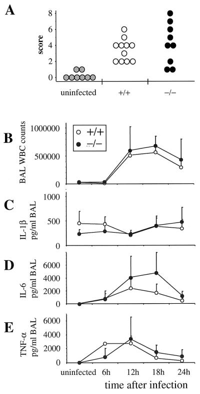
FIG. 3.
Histopathology and inflammatory response are unchanged in mBD-1−/− mice. (A) Histopathological assessment of lung sections obtained from uninfected mice and from mBD-1+/+ and mBD-1−/− animals 24 h after infection with 107 CFU of H. influenzae. Individual sections were scored in a blinded fashion for inflammatory responses, which predominantly consisted of neutrophilic infiltration around airways and into the alveolar space. (B to E) Kinetics of the inflammatory response to infection with 5 × 10⁷ CFU H. influenzae. BAL fluid was collected at the indicated time points, and total BAL white cell (WBC) counts were determined (B), as well as levels of IL-1β (C), IL-6 (D), and TNF-α (E). Three animals per group were analyzed per time point, except for 12 h (11 mBD-1+/+ and 9 mBD-1−/− animals in each group) and 24 h (6 animals in each group).

