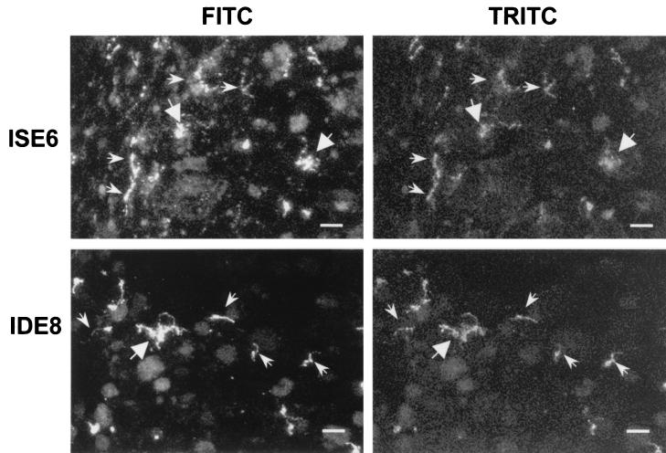
FIG. 2.
Extracellular location of B. burgdorferi N40 in infected tick cell cultures. B. burgdorferi (MOI, 5) was cultured for 3 days with IDE8 or ISE6 tick cells. Confocal integrated micrographs of B. burgdorferi in unpermeabilized and permeabilized tick cell cultures show similar spirochetal staining with each reagent. The small arrows indicate single B. burgdorferi cells, and the large arrows indicate large clumps of B. burgdorferi cells. Bars, 100 μm.

