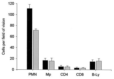
FIG. 4.
Mean values and standard deviations for number of cells per field of vision (magnification, ×400) for mice infected with cytotoxin-positive (solid bars) (n = 22) and cytotoxin-negative (shaded bars) (n = 21) H. pylori strains. Gastric mucosa in mice infected with cytotoxin-positive H. pylori strains showed significantly more polymorphonuclear leukocytes than gastric mucosa in mice infected with cytotoxin-negative H. pylori-strains (P < 0.0001). No differences was detected for any other cell type analyzed.

