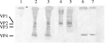
FIG. 1.
Autoradiogram of CVB capsid polypeptides on 16% sodium dodecyl sulfate-polyacrylamide gel electrophoresis under reducing conditions. Virus polypeptides VP1, VP2, VP3, and VP4 are indicated by lines on the left. Lane 1, mock virus obtained from supernatant of noninfected Hep-2 cells; lanes 2 and 3, 35S-labeled CVB3 and 35S-labeled CVB4E2 from CVB3- and CVB4E2-infected Hep-2 cells, respectively; lanes 4 and 5, H antigen from 35S-labeled CVB3 and 35S-labeled CVB4, respectively, dissociated at 56°C as described in Materials and Methods; lanes 6 and 7, VP4 from 35S-labeled CVB3 and 35S-labeled CVB4E2, respectively, obtained after dissociation.

