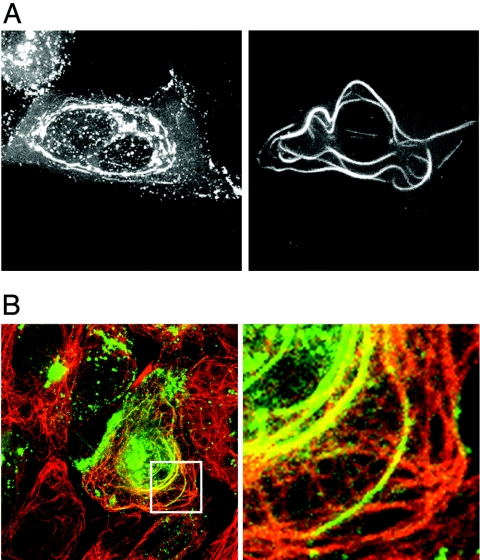
FIG. 4.
The G22P− protein forms MT bundles in infected cells. Vero cells infected with G22P-v at a multiplicity of infection of 0.1 were examined live at 20 h postinfection (A) or were fixed with methanol and processed for immunofluorescence with an anti-α-tubulin antibody. (B) Cells were examined by confocal microscopy for GFP-VP22 (green) and α-tubulin fluorescence (red). Right-hand panels show magnified images of the region in the white box.

