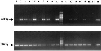
FIG. 5.
RT-PCR analysis of IDO mRNA expression in dTHP-1 cells and MdM stimulated for 24 h with C. trachomatis-infected HeLa cell supernatants and in dTHP-1 cells cocultivated with infected HeLa cells for 24 and 48 h. The cDNAs were amplified with IDO (324 bp, top panel) and GAPDH (306 bp, bottom panel) primers for 35 and 22 PCR cycles, respectively. A total of 10 μl of PCR products was loaded on a 2% agarose gel as follows. dTHP-1 cells and MdM were incubated with supernatants from HeLa cells left uninfected (lanes 1 and 6, respectively), infected with serovar E (lanes 2 and 7, respectively) or serovar L2 (lanes 3 and 8, respectively), treated with RPMI alone (lanes 4 and 9, respectively) or E. coli LPS alone (lanes 5 and 10, respectively), or dTHP-1 cells were incubated in coculture for 24 and 48 h with uninfected HeLa cells (lanes 11 and 14, respectively), with serovar E-infected HeLa cells (lanes 12 and 15, respectively), or with serovar L2-infected HeLa cells (lanes 13 and 16, respectively). A negative control for amplification (lane 17) wherein DNA was omitted and a positive control (lane 18) consisting of cDNA from HeLa cells exposed to rhIFN-γ (10 ng/ml for 12 h) were also included in the analysis.

