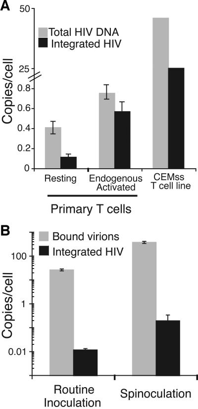
FIG. 2.
HIV-1 integrated into resting CD4+ T cells. (A) The relative levels of total DNA and integrated DNA in the two populations of primary T cells at 3 days postinoculation are shown. Total DNA was measured by primers that detect second strand transfer. Integrated DNA was measured by Alu-PCR (51). The relative levels of total DNA and integrated DNA for the activated T-cell line, CEMss, at 18 h postinoculation are also shown. Immediately after spinoculation only very low levels of total DNA (<0.001 copies/cell) and no integrated DNA were detected (not shown), indicating that our viral supernatants contained minimal amounts of contaminating viral DNA from the initial transfection. Comparing the resting and endogenous activated CD4+ T cells, there was a significant difference in the amount of total DNA (P < 0.02) and integrated DNA (P ≪ 0.01) by Student's t test. Similar results were obtained with HIV-1YU2, a CCR5-tropic virus, and pNL4-3, another CXCR4-tropic virus (not shown). The data are representative of four experiments. (B) Spinoculation did not induce integration. Binding and integration were increased proportionally by spinoculation. Resting CD4+ T cells were inoculated under routine and spinoculation conditions and washed to remove unbound virions, and the number of bound virions per cell was measured as described elsewhere (52). The number of integrated proviruses per cell was measured 3 days after inoculation as described previously (51). The data are representative of three experiments.

