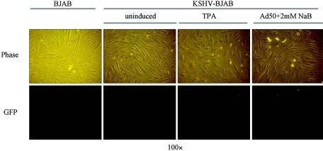
FIG. 5.
Recombinant KSHV virus reconstituted from induced KSHV-BJAB cells can be transmitted to hTERT-immortalized microvascular endothelial (TIME) cells. Phase (upper panel) and eGFP fluorescence (lower panel) are shown. Three days postinduction, supernatant was briefly centrifuged to remove cell debris and then filtered through a 0.45-μm-pore filter. The resulting supernatant was used to infect TIME cells on a 12-well plate. Twenty-four hours postinfection, phase and fluorescence pictures were taken. Supernatant from BJAB cells was used to infect TIME cells (left panel) as a negative control. TIME cells infected with supernatants from uninduced (middle-left panel), TPA-induced (middle-right panel) and Adeno-50 (Ad50) plus 2 mM sodium butyrate (NaB)-induced (right panel) KSHV-BJAB cells are shown. Quantification is described in the text.

