FIG. 4.
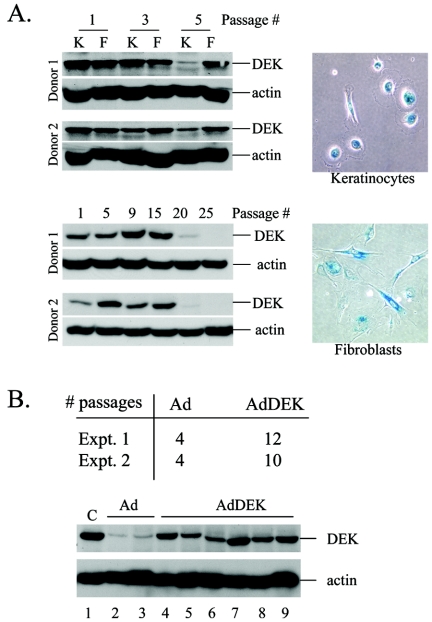
DEK expression is decreased during replicative senescence of human cells, and ectopic DEK expression extends keratinocyte life span. (A) Keratinocyte (K) and fibroblast (F) cultures were prepared from human foreskin tissue. Protein lysates were prepared at the indicated passage numbers. Equal amounts of total protein were subjected to DEK-specific Western blot analysis. Keratinocytes at passage 5 and fibroblasts at passage 25, respectively, were stained for SA-β-Gal activity and photographed. (B) Keratinocytes were infected with empty Ad or AdDEK at an MOI of 100 and were split when they reached approximately 60 to 80% confluence. For experiment 1, the cells were split 1:4 at every passage and infected twice at passages 3 and 4. For experiment 2, the cells were split to 5 × 10⁵ cells per 10-cm plate at every passage and infected after each split, starting at passage 3. Passage numbers were recorded. Western blot analysis for experiment 2 is shown underneath. A portion of the cells was removed at every split and was subjected to DEK-specific Western blot analysis.

